
Translational research often commences in various models – cells, rodents, slice cultures.. But results from all of these rest on an 𝙖𝙨𝙨𝙪𝙢𝙥𝙩𝙞𝙤𝙣 that the underlying cause of the human disease is correctly replicated in the chosen model.
There is no single easy way to prove this assumption, especially in a complex and often multi-factorial condition like Parkinson’s disease. One method that is available to us is the use of donor tissue from people who received the relevant diagnosis. Brain tissues are collected postmortem (following informed consent obtained during life), and findings on the cellular level in the brain can be then correlated with symptoms and other features.
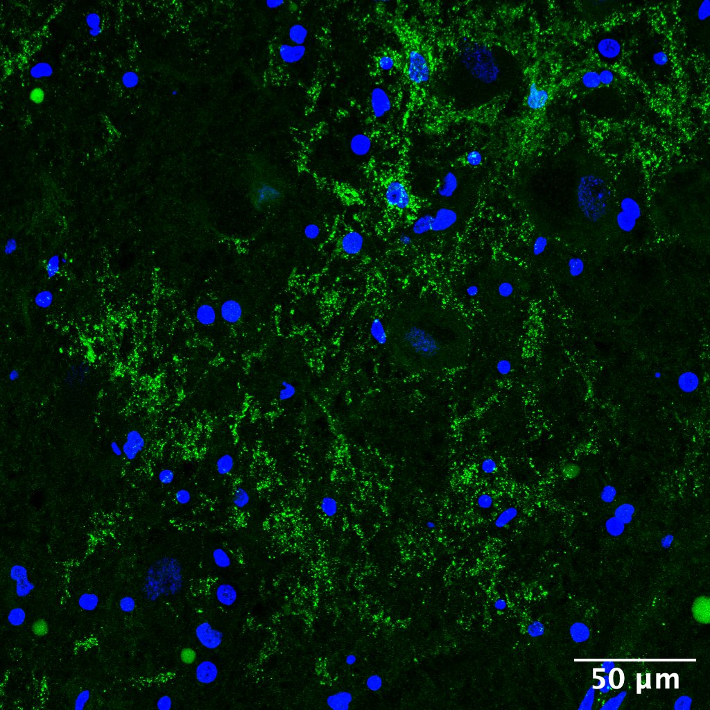
Analysis of 20 late-stage Parkinson’s cases compared with 20 age-matched controls revealed dysregulation in an astrocytic network-forming protein Connexin43 which was associated with Parkinson’s:
https://www.nature.com/articles/s41598-025-94188-7
The full dataset can be explored using our interactive web app: https://www.cx43pd.com
These findings give us a clue to look for this specific astrocytic pathology in relevant preclinical models, and ultimately find a way to safely protect astrocytic networks with an aim of minimising symptom progression.
Special thanks to Nature Portfolio for publication, Parkinson’s UK Brain Bank for the support with the tissue sourcing, Cambridge Stem Cell Institute for the access to the excellent imaging facility, and Rosetrees for providing the Seedcorn grant that kick-started this project.

Image: an astrocyte in the human frontal cortex “holding” a blood vessel
(Dr Nataly Hastings, Dr Saifur Rahman, and Dr P. Aleks Stempor are co-authors of the article and members of the founding team at Cellestial Health. The data presented in the article are entirely the result of the prior collaborative work at the University of Cambridge sponsored by academic grants.)